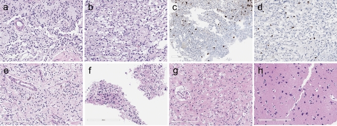
Figure 2.
Pathological features of DNET (D01) at the first and second operations. The patient underwent two operations due to tumor recurrence. (a) and (b) The main tumor (M1) and a satellite nodule (SL1) at the first operation show the same histology composed of oligodendrocyte-like cells (OLCs) in the myxoid background. (c) and (d) NeuN-positive floating neurons are small and are hardly seen in DNET (a, c M1; b, d SL1). In the recurrent tumor, (e) the main tumor (rM3) and (f) a large SL (rSL1) show the same appearance as typical DNETs, which are composed of monotonous OLCs with floating neurons. The other, smaller SLs, (g) rSL3 and (h) rSL5 are not tumors, but contain reactive gliosis and foamy macrophage infiltration. (a, b, e–h H & E stain; c, d NeuN immunohistochemistry; Scale bar: 200 μm).

