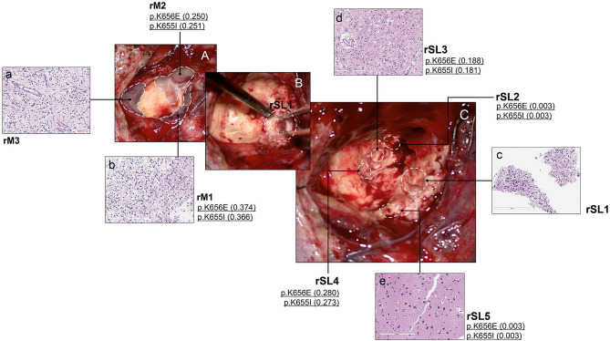
Figure 3.
Intraoperative photographs and histological/genetic exams of Case D01 at the second operation. The patient developed multifocal recurrence in the previous tumor cavity along the resection margin (A). The main tumor was divided into 3 parts (lateral (rM1), anterior (rM2), and medial (rM3)). After removal of the main masses, hidden satellite lesions (SLs) were further searched for and removed as much as possible. SL1 was most conspicuous having formed a discrete mass (B) and the others (SL2-5) were smaller, consisting of tiny nodules. The locations of SLs are marked by dotted circles after the completion of tumor resection (C). If a tumor sample was sequenced, the presence of FGFR1 mutation and VAF is described. Both medial and lateral masses show the same histology composed of OLCs in the myxoid background with the small number of floating neurons, consistent with DNET (a, b). SL1 has similar tumor histology, filled with OLCs and floating neurons (c). WES revealed mutations in the FGFR1 gene. SL2 and SL4 were too small, and only WES was performed without histologic examinations, which revealed the same FGFR1 mutations as SL1 had. In SL3 and SL5, histologic examinations reveal only gliosis and foamy macrophage infiltration without obvious DNET-like features, but FGFR1 mutations are also found in SL3 and SL5 (d, e).

