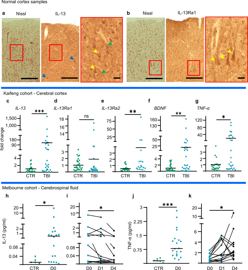
Fig. 9. IL-13 is expressed in the human brain and is upregulated upon TBI in the human cortex and CSF.
a Darrow red Pigment-Nissl and Immunohistochemistry for IL-13 shows moderate, high and very high-expressing neuronal populations in human post-mortem cortical tissue. N = 3. b Darrow red Pigment-Nissl and Immunohistochemical staining of IL-13Ra1 shows a large number of neurons across all cortical layers of post-mortem cortical tissue. N = 3. a, b Scale bar overview: 100 μm, scale bar insert: 10 μm. c–g Significant upregulation of the mRNA for IL-13 (p = 0.0002), IL-13Ra2 (p = 0.0021), BDNF (p = 0.0015) and TNF-α (p = 0.0160), but not of IL-13Ra1 (p = 0.0998), in human cortical tissue samples resected after traumatic brain injury vs controls from elective surgery. (RT-qPCR). CTR N = 34; TBI N = 29. h Significant upregulation of IL-13 protein in cerebrospinal fluid samples from severe TBI patients within 24 h of TBI (p = 0.0243). CTR N = 5; TBI N = 18. i Time course of CSF levels of IL-13 in severe TBI patients: progressive decrease of IL-13 levels between D0 and D4 (p = 0.0016). D0 N = 12; D1 N = 12; D4 N = 12. j Significant upregulation of TNF- α in CSF of TBI patients (p < 0.0001). CTR N = 5; TBI N = 20. k Progressive increase of TNF-α levels in the CSF of severe TBI patients between d0 and D4 (p = 0.0181). D0 N = 12; D1 N = 12; D4 N = 12.Cytokines measured by SIMOA assay. *: p < 0.05. **: p < 0.01. ***: p < 0.001. c–g, h, j Two-tailed Mann–Whitney test. i, k Friedman test with Dunn’s multiple comparison. Source data are provided as a Source Data file.

