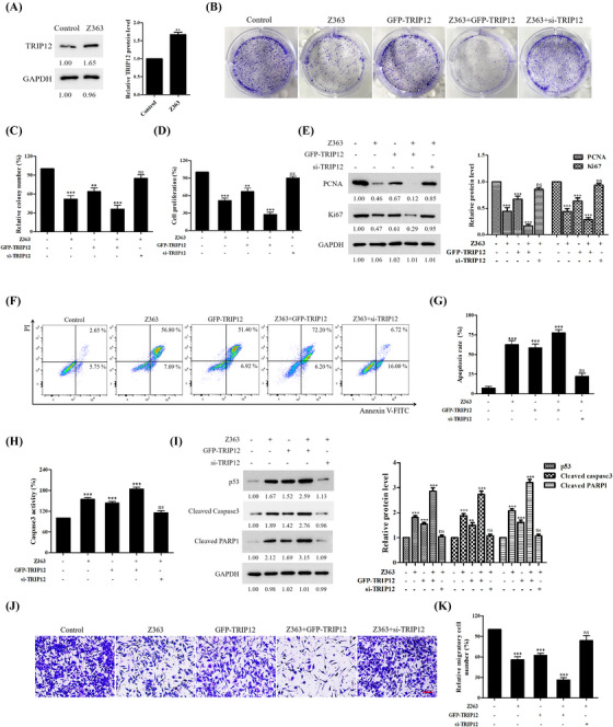
FIGURE 5.

TRIP12 inhibits breast cancer cells proliferation, metastasis and induces apoptosis. (A) MCF7 cells were treated with Z363 (7.5 μg/ml) for 24 h. The protein levels of TRIP12 were analysed by Western blotting. (B) MCF7 cells were transfected with GFP‐TRIP12 or si‐TRIP12 for 24 h, followed by treated with or without Z363 (7.5 μg/ml) for 24 h. The ability of cell to form colonies was measured using crystal violet staining. (C) Statistical analysis of colony formation. (D) MCF7 cells were transfected with GFP‐TRIP12 or si‐TRIP12 for 24 h, followed by treated with or without Z363 (7.5 μg/ml) for 24 h. Cell proliferation of groups were measured by a Ki67 ELISA kit. (E) The expressions of Ki67 and PCNA were analysed by Western blotting. (F) MCF7 cells were transfected with GFP‐TRIP12 or si‐TRIP12 for 24 h, followed by treated with or without Z363 (7.5 μg/ml) for 24 h. MCF7 cells were treated with different groups, followed by assessment of apoptosis by flow cytometry analysis. (G) Statistical analysis of cell apoptosis. (H) Caspase‐3 activity was measured in MCF7 cells treated with different groups. (I) The expressions of Cyto C and p53 were analysed by Western blotting. (J) Cells were transfected with GFP‐TRIP12 or si‐TRIP12 for 24 h, followed by treated with or without Z363 (7.5 μg/ml) for 24 h. Cell migration ability of groups were measured by transwell assays. Scale bar, 20 μm. (K) Statistical analysis of migration ability. Data shown in C, D and G, H, K were analysed by one‐way ANOVA. Colony images, fluorescence images, transwell and blots were representative of three independent experiments. All data are presented as the mean ± SEM of n = 3. ***p < .001, **p < .01, ns, no significance
