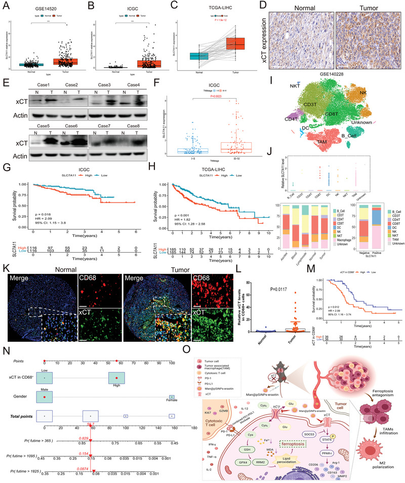
Figure 9.

Distinctive clinical functional profiles of xCT and macrophage‐derived xCT in HCC. A–C) Expression of SLC7A11 in tumor tissues and normal tissues in GSE14520 (A), ICGC (B), and TCGA‐LIHC (C). D) IHC staining showing the differences in the expression of xCT in tumor tissue and normal tissue. E) Western blot exhibiting the expression characteristics of xCT in tumor tissues and normal tissues. F) The correlation between SLC7A11 levels and TNM staging in ICGC database. G,H) Survival analysis showing the predictive value of SLC7A11 in survival probability in ICGC (G) and TCGA‐LIHC cohort (H). I) The immune cellular landscape of HCC. J) Expression of SLC7A11 in immune cells and the distribution proportion of immune cells in different tissues. K,L) IF staining showing the proportion of infiltrating CD68+ cells and xCT expression in CD68+ cells in tumor tissues compared to normal tissues. M) Survival analysis indicating a significant survival advantage of HCC patients with low xCT expression in CD68+ cells. N) A nomogram was constructed based on the independent prognostic factors of HCC. O) The overall process and mechanism of this study. Created with Biorender.com. Data in the figure are represented as the means ± SEM. Differences between the groups were evaluated using Student's t‐test. *** p < 0.001.
