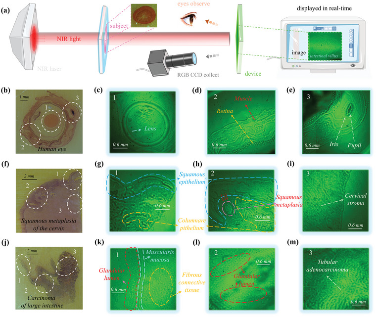
Figure 3.

Bioimaging applications of the NIR‐OPD. a) Schematic diagram of bio‐imaging process and method by utilizing our NIR‐OPR and imaging results of small intestine (on the right of the annotation “image”). b) Images of transection of human eye sample directly collected by RGB CCD and c–e) imaging under 850 nm NIR laser. f) Images of squamous metaplasia of cervix sample directly collected by RGB CCD and g–i) imaging under 850 nm NIR laser. j) Images of carcinoma of large intestine sample directly collected by RGB CCD and k–m) imaging under 850 nm NIR laser.
