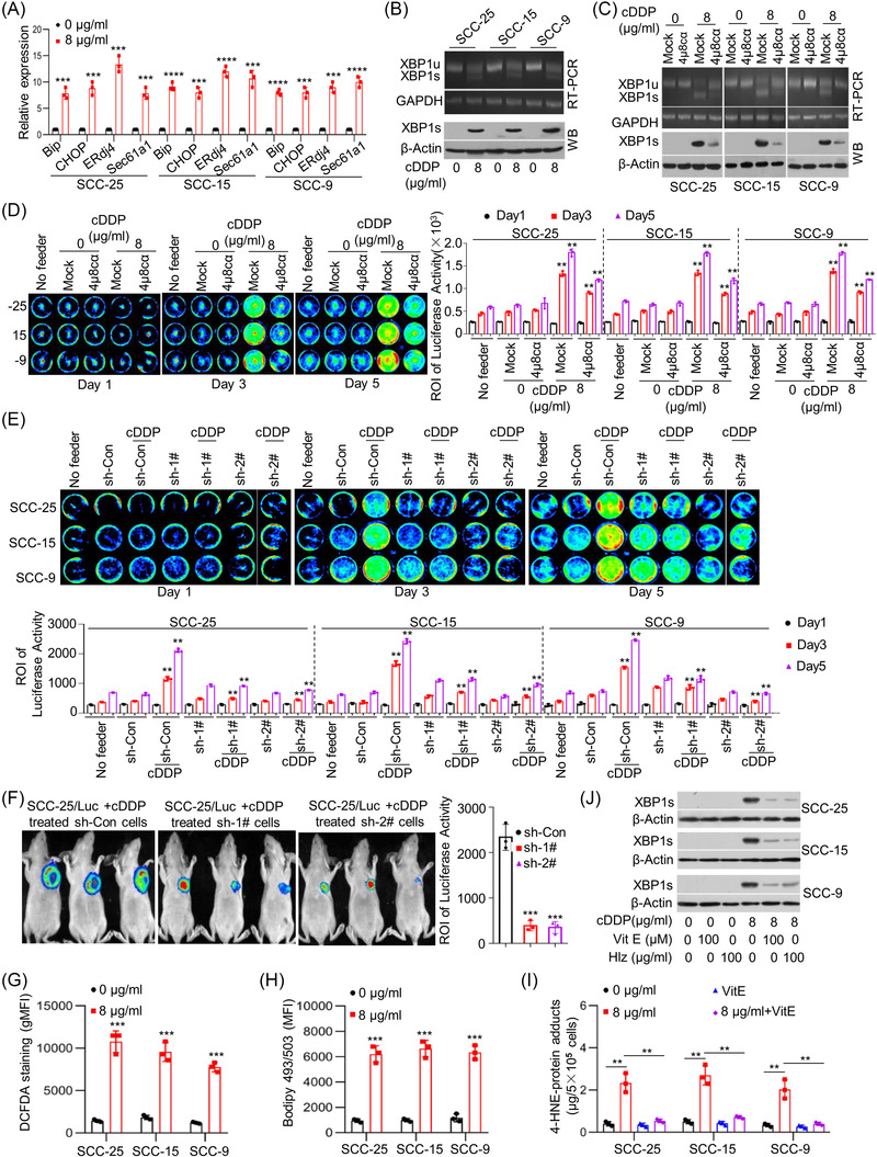
FIGURE 2.

Cytotoxic treatment‐induced endoplasmic reticulum (ER) stress, mediating tumour cell repopulation. (A) SCC‐25, SCC‐15 and SCC‐9 cells were treated with cisplatin (cDDP) at the indicated concentrations for 24 h. The expression levels of BiP, CHOP, ERdj4 and Sec61a1 were measured by quantitative real‐time polymerase chain reaction (qRT‐PCR) (n = 3). *** p < .001, **** p < .0001. (B) X‐box binding protein‐1 (XBP1) splicing was evaluated using RT‐PCR (upper) and western blots (bottom). (C) SCC‐25, SCC‐15 and SCC‐9 cells were treated with vehicle (mock) or the IRE1α inhibitor 4µ8cα (10 µM) in combination with cDDP for 24 h. XBP1 splicing was evaluated using RT‐PCR (upper) and western blots (bottom). (D) SCC‐25/Luc, SCC‐15/Luc and SCC‐9/Luc reporter cells were seeded among the respective feeder cells with or without treatment with the IRE1α inhibitor 4µ8cα in combination with cDDP or alone in 24‐well plates. Cancer cell repopulation in vitro was observed by luciferase activities. ** p < .01. (E) SCC‐25, SCC‐15 and SCC‐9 cells with or without XBP1 knockdown were treated via cDDP for 24 h in 24‐well plates. Then, SCC‐25/Luc, SCC‐15/Luc and SCC‐9/Luc reporter cells were seeded among the respective feeder cells. Cancer cell repopulation in vitro was measured by luciferase activities. ** p < .01. (F) SCC‐25 cells with XBP1 knockdown or control vector were treated via cDDP for 24 h. Then, SCC‐25/Luc cells were injected subcutaneously together with above cells into nude mice, and the repopulation of SCC‐25/Luc cells was represented by luciferase levels, *** p < .001. (G) The intracellular reactive oxygen species (ROS) levels in SCC‐25, SCC‐15 and SCC‐9 cells treated with cDDP were evaluated using 2′,7′‐dichlorofluorescin diacetate (DCFDA) staining (n = 3), *** p < .001. (H) The intracellular lipid content in SCC‐25, SCC‐15 and SCC‐9 cells treated with cDDP was quantified using 4,4‐difluoro‐1,3,5,7,8‐pentamethyl‐4‐bora‐3a,4a‐diaza‐s‐indacene (BODIPY 493/503) staining (n = 3), *** p < .001. (I) The intracellular levels of 4‐hydroxy‐trans‐2‐nonenal (4‐HNE)‐protein adducts in SCC‐25, SCC‐15 and SCC‐9 cells treated with cDDP in combination with ROS‐scavenging agent vitamin E (VitE) using enzyme‐linked immunosorbent assays (ELISAs) (n = 3), ** p < .01. (J) SCC‐25, SCC‐15 and SCC‐9 cells were treated with cDDP in combination with VitE (100 µM) or 100 µg/ml hydralazine (Hlz). XBP1 splicing was detected by western blotting. Data in (A) and (D)–(I) are represented as the mean ± SEM. Statistical significance was determined by a two‐tailed Student's t‐test
