Fig. 1.
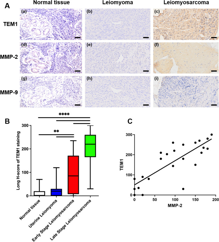
The expression of TEM1, MMP-2 and MMP-9 in different uterine samples. A Representative immunohistochemical results of TEM1, MMP-2 and MMP-9 staining. TEM1 and MMP-2 were negative in uterine myometrium (normal tissue) and leiomyoma, strong positive in leiomyosarcoma; MMP-9 staining was negative or weakly expressed in all samples. Scale bar, 50 μm. B Boxplot for Long H-scores of TEM1 in normal myometrium, leiomyoma and leiomyosarcoma tissues. TEM1 Long H-scores were positively correlated with tumor stages, r = 0.9554. **, P < 0.01; ****, P < 0.0001. C TEM1 expression was positively correlated with MMP-2 expression in uterine leiomyosarcoma tissues. Correlation curves are shown with r square, and P‐values from Pearson´s correlation analysis. R square = 0.6487, Pearson r = 0.8054, P < 0.0001

