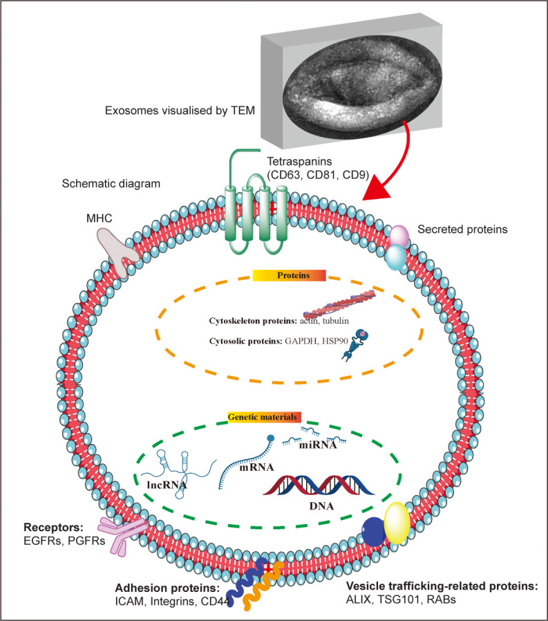
Figure 1. Exosome morphology and structure. The upper image represents the morphology of exosomes (produced by mouse hepatocytes) under a transmission electron microscope; below is a schematic diagram of the structure and composition of exosomes. ALIX: apoptosis-linked gene 2-interacting protein X; EGFR: epidermal growth factor receptor; GAPDH: glyceraldehyde 3-phosphate dehydrogenase; HSP90: heat shock protein 90; ICAM: intercellular adhesion molecule; lncRNA: long non-coding RNA; MHC: major histocompatibility complex; miRNA: microRNA; PGFR: platelet-derived growth factor receptor; RAB: Rab GTPases; TEM: transmission electron microscope; TSG101: tumour susceptibility gene 101. This figure was created using Servier Medical Art templates (https://smart.servier.com).

