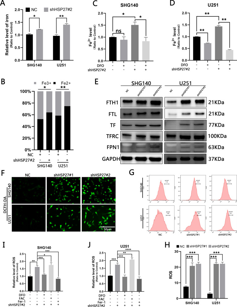
Fig. 3.
Silencing HSP27 increases cellular ferrous ion-mediated ROS production. A, B Total iron levels and ferrous ion ratios before and after shHSP27 transfection of SHG140 and U251 cell lines were determined by colorimetric methods. C, D Colorimetric determination of ferrous ion expression in different groups after DFO treatment. E Expression of iron-associated proteins in control groups and after lentiviral transfection. F Observation of ROS fluorescence intensity before and after transfection under fluorescence microscopy, DCFH-DA staining, scale = 50 μm. G, H Flow cytometry quantitatively detects ROS before and after transfection. I, J The microplate reader detects changes in ROS in different treatment and control groups. The statistical analysis showed *P < 0.05, **P < 0.01, ***P < 0.001, ****P < 0.0001, NSP > 0.05

