Figure 3.
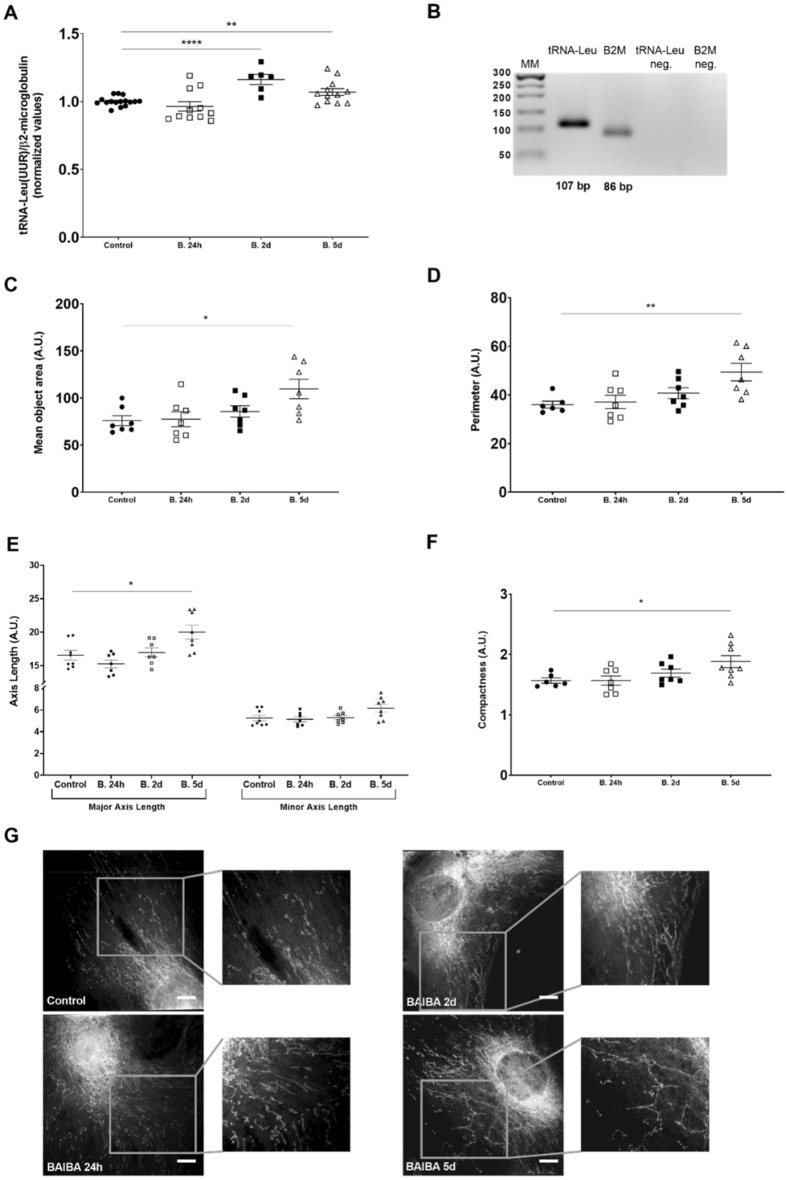
L-BAIBA promotes mitochondrial enlargement and branching in podocytes. (A) L-BAIBA treatment (10 µM; 24 h, 2 days, and 5 days) influenced mtDNA levels in podocytes. n = 6–16. **p < 0.01, ****p < 0.0001. (B) Representative agarose gel showing PCR products. (C) Mean mitochondrial area (total number of pixels in the object) in podocytes after L-BAIBA treatment. n = 7. *p < 0.05. (D) Mitochondrial perimeter (number of pixels that formed the boundary of an object) in podocytes after L-BAIBA treatment. n = 6–7. **p < 0.01. (E) L-BAIBA affected major axis length (pixel distance between endpoints on the longest line that could be drawn through the object) and minor axis length (longest line that could be drawn through the object while remaining perpendicular to the major axis) of mitochondria in podocytes. n = 7–8. *p < 0.05. (F) L-BAIBA affected mitochondrial compactness ([perimeter]2 / [4π × area]). n = 6–8. *p < 0.05. (G) Confocal images of podocyte mitochondria after L-BAIBA treatment. Scale bar = 10 μm.

