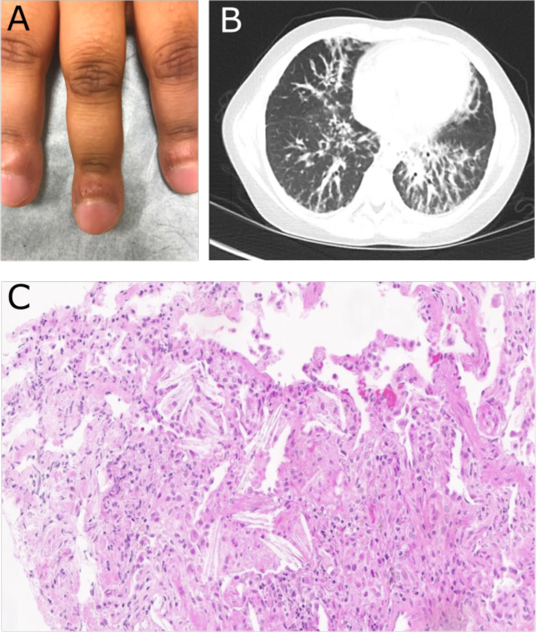
Figure 1: Physical exam, radiographic, and histologic markers of SJIA-LD.

(A) Photograph of fingers prior to MAS-825 showing significant clubbing and erythematous, inflamed periungual tissue. (B) Representative image of lung CT prior to MAS-825 therapy demonstrating peribronchovascular interstitial thickening. (C) Photomicrograph of transbronchial biopsy of lesion demonstrating interalveolar cholesterol clefts (lower right) and foamy macrophages (upper left) consistent with the endogenous lipoid pneumonia pattern of SJIA-LD.
