
Figure 4: Photomicrograph of cytospin from BAL prior to MAS-825 therapy.
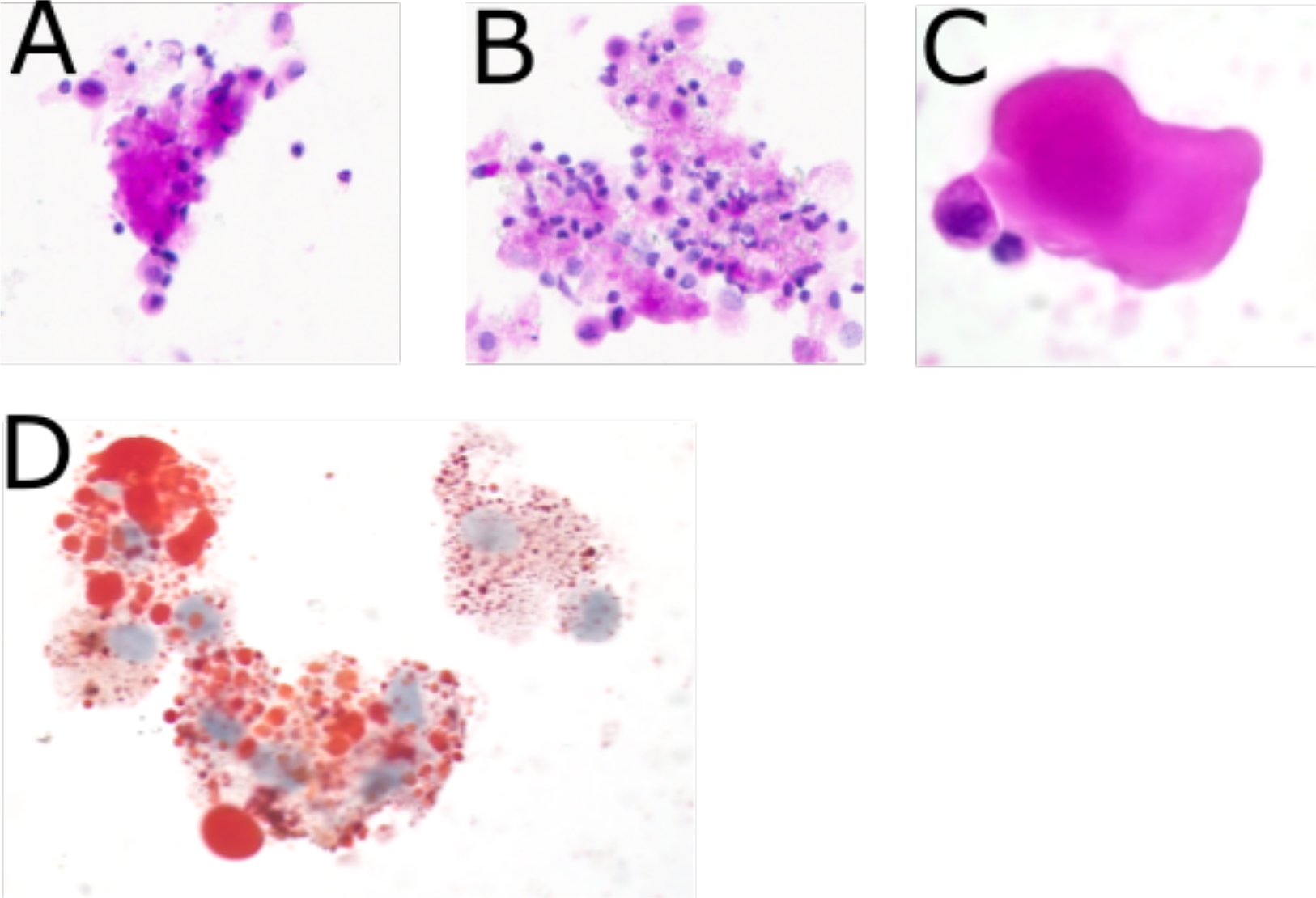
(A) Acellular protein globule consistent with PAP. (PAS stain, 40X) (B) Numerous foamy macrophages present within the fluid. (PAS stain 40X) (C) High power view of acellular protein globule. (H&E stain 100X) (D) Lipid laden foamy macrophages. (Oil Red O stain 100X)