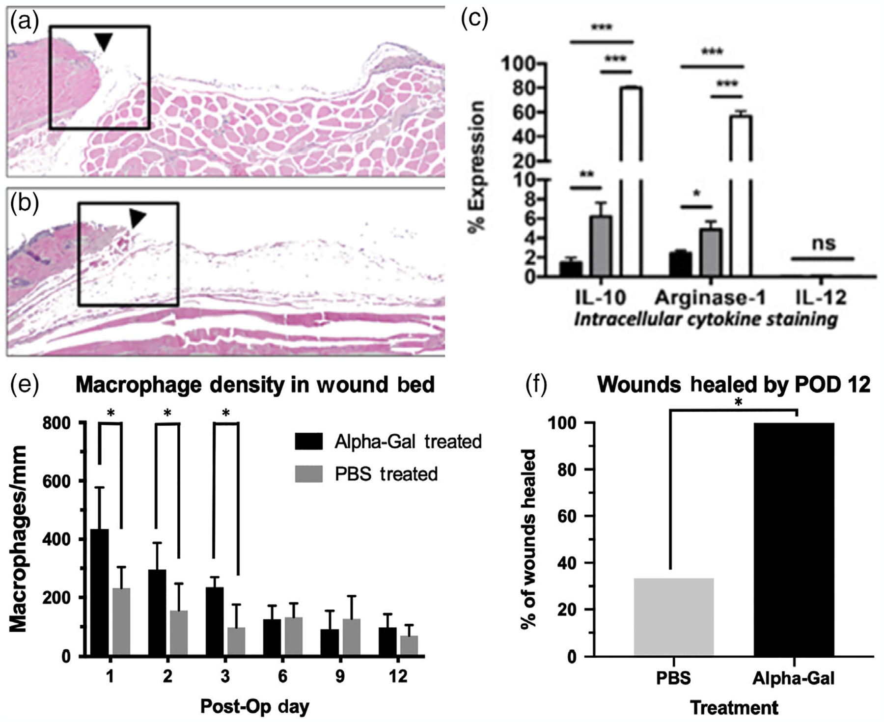
FIGURE 9.

α-Gal epitope expressing nanoparticles for the recruitment of macrophages for wound healing treatment. (a,b) H&E longitudinal hemi section of wounds showing macrophage density within wounds (a: Treated vs. b: Untreated). (c) Flow cytometry data. Closed column: PMN, gray columns: Medium size macrophages, open columns: Large size macrophages. Most macrophages are M2. (d) Macrophage density in wound bed. Greater density was observed in AGN (α-gel)-treated wounds. (e) Healing rate showing 100% healing by POD12 versus 33% in PBS (untreated) wounds (Kaymakcalan et al., 2020).
