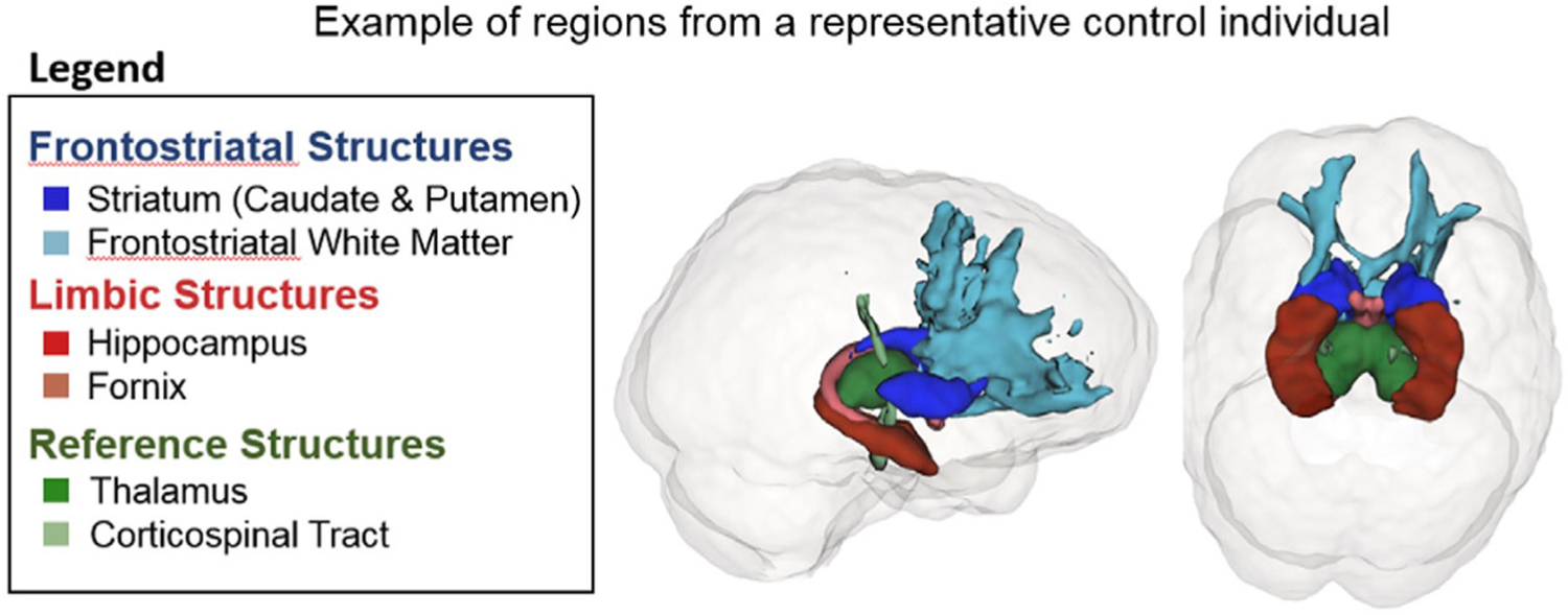
FIGURE 2.

Representation of frontostriatal, limbic, and reference structures. Representative image of regions studied: striatum (dark blue), frontostriatal white matter (light blue), hippocampus (dark red), fornix (light red), thalamus (dark green), and corticospinal tract (green)
