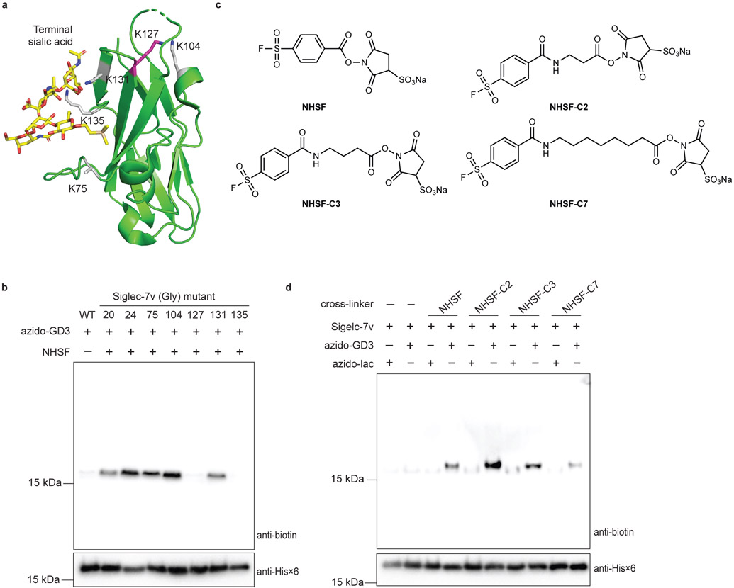
Fig. 3. Cross-linking site on Siglec-7v and distance dependence of the cross-linker indicate that sulfonyl fluoride of NHSF reacted with carbohydrate via proximity-enabled reactivity.
a) Crystal structure of Siglec-7v binding with α(2,8)-disialygangioside GT1b (PDB: 2HRL). NHSF cross-linking site, Lys127, on Siglec-7v is shown in magenta stick. All other Lys sites are shown in grey stick. b) NHSF cross-linking of azido-GD3 with Siglec-7v Lys to Gly mutants. c) Structures of NHSF analogs with different linker lengths. d) Cross-linking of Siglec-7v with azido-GD3 by the NHSF analogs. Faint background bands in the anti-biotin blots were due to low level reaction of alkyne-biotin with protein Siglec-7v nonspecifically, a common background when using azide-alkyne for click labeling as previously reported.36,37

