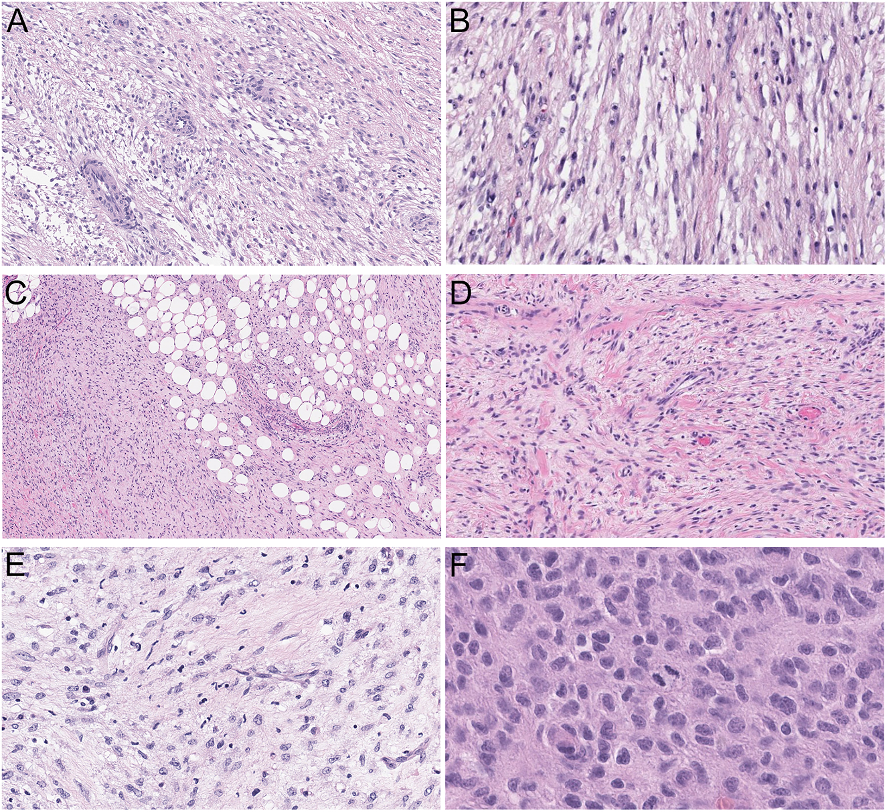
Figure 2. Histologic morphology.

A-E, Low-grade fibromyxoid tumors consisting of fusiform cells arranged in loosely formed fascicles set in a collagenous to myxoid stroma (A-B: case 2, 100X and 200X; C-D: case 1, 100X and 200X; E: case 3). A, D, Admixed thin-walled, round to linear vasculature. B, E, Occasional lymphocytes and neutrophils admixed with tumor cells. C, Focal honeycombing of fat, reminiscent of dermatofibrosarcoma protuberans. F, The metastatic tumor of case 3 displayed a more prominent epithelioid morphology with irregular, eccentric nuclei and moderate amount of eosinophilic cytoplasm with increased mitotic activity (400X).
