
Figure 1:
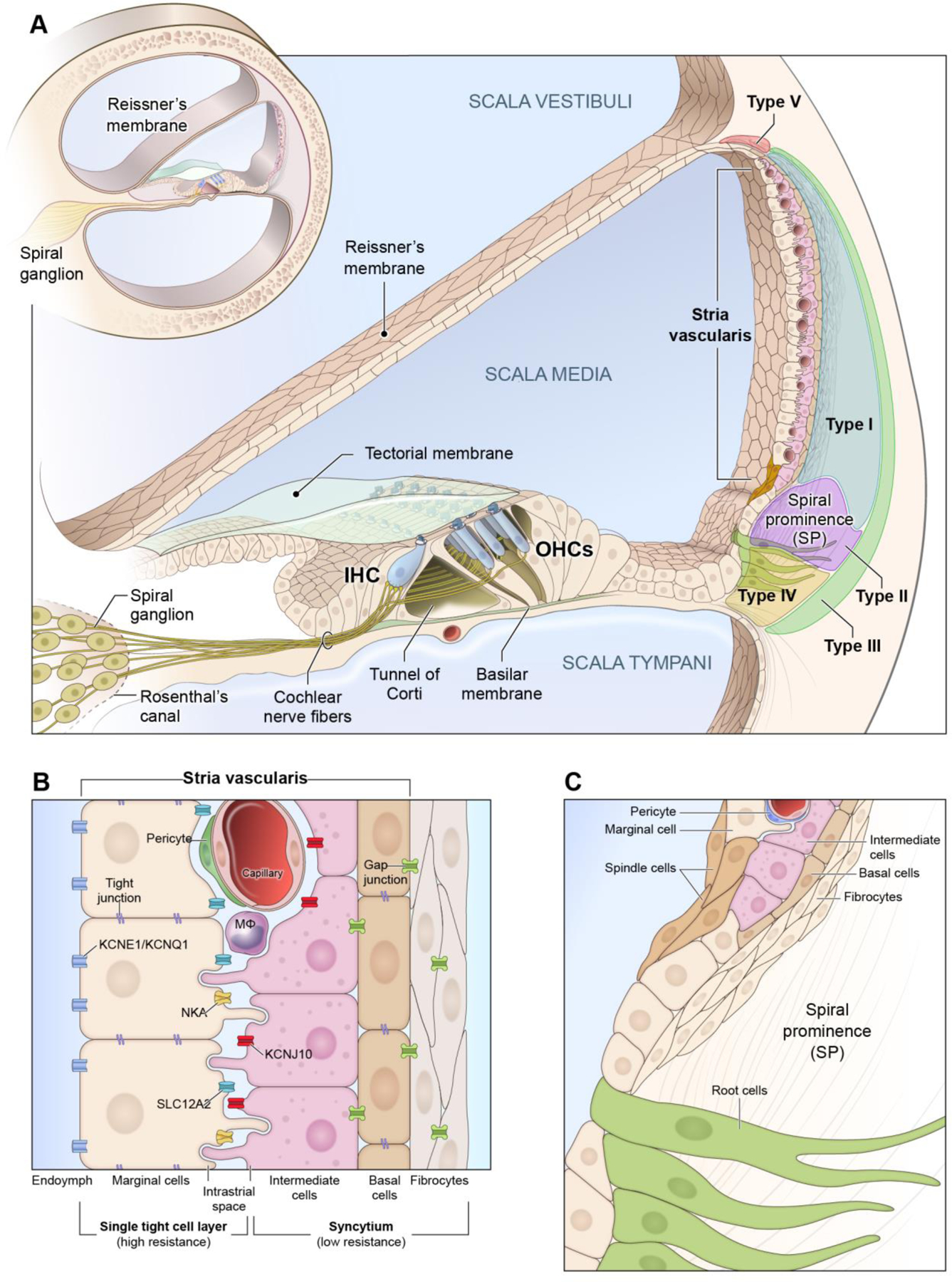
Schematic of Stria Vascularis (SV)
A, The mammalian cochlea consists of 3 fluid-filled chambers with two chambers (scala tympani and scala vestibuli) containing perilymph and one chamber (scala media) containing endolymph. The SV is housed in the lateral wall of the cochlea and its 3 main cell layers are composed of marginal cells (MC), intermediate cells (IC), and basal cells (BC), respectively, with the marginal cells facing the endolymph. The SV generates the highly positive endocochlear potential (+80 mV or greater) and is responsible for the high potassium concentration of the endolymph which distinguishes it from the perilymph in the other two fluid-filled chambers. Fibrocyte populations in the spiral ligament (Types I-V) are also depicted. B, Magnified view of the layers of the stria vascularis with major ion channels implicated in its function. Marginal cells excrete potassium into the endolymph and accomplish this process through their expression of a group of ion channels, including KCNE1/KCNQ1, SLC12A2, and NKA. Marginal cells are connected by tight junctions apically. Marginal cells extend processes basally toward the intermediate cells which also extend processes apically toward the marginal cells and basally towards the basal cells. Macrophages, pericytes, and endothelial cells form the basis for the blood-labyrinth barrier (BLB). The space between the marginal and intermediate cells forms the intrastrial space which has a low potassium and the inward-rectifier K+ channel, KCNJ10, concentrates potassium in the intermediate cells and is critical to the ability of the stria vascularis to both generate the endocochlear potential (EP) and regulate cochlear ionic homeostasis in the endolymph. Both basal and intermediate cells expression gap junctions. Basal cells along with intermediate cells and fibrocytes function together as a syncytium and are connected through gap junctions and maintain a barrier through a network of tight junctions. C, Rare spindle and root cells are located at the superior and inferior periphery of the stria vascularis and the region adjacent to the spindle cells, respectively. Inferiorly, the spindle cells overlie the spiral prominence and extend superiorly toward the edge of the stria vascularis, contacting marginal cells. Spindle cells and root cells both are known to express SLC26A4 (otherwise known as pendrin) and KCNJ16 (Korrapati, Taukulis et al., 2019; Gu et al., 2020). Abbreviations: Na+-K+-ATPase (NKA), Na+/K+/Cl− symporter 1 (SLC12A2), potassium voltage-gated channel subfamily E regulatory subunit 1 (KCNE1), potassium voltage-gated channel subfamily Q regulatory subunit 1 (KCNQ1), ATP-sensitive inward-rectifier potassium channel 10 (KCNJ10), solute carrier family 26 member 4 (SLC26A4) otherwise known as pendrin, potassium inwardly rectifying channel subfamily J member 16 (KCNJ16).