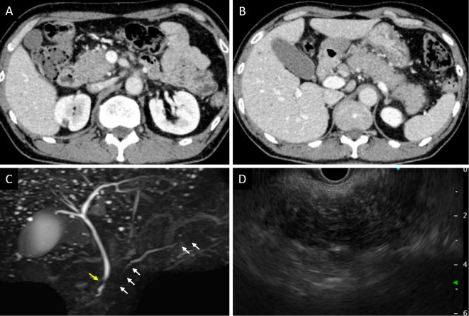
Figure 2.
Imaging findings of the pancreas. CECT showed diffuse enlargement of the pancreas, peripancreatic fat stranding, no acute peripancreatic fluid collection, and no capsule-like rim in the head of the pancreas (A) or from the body of the pancreas to the tail (B). MRCP showed irregular narrowing of the main pancreatic duct in the head and tail (white arrows) and localized stricture of the distal common bile duct (yellow arrow) (C). On EUS, a diffusely hypoechoic and enlarged pancreas was observed (D). CECT: contrast-enhanced computed tomography, MRCP: magnetic resonance cholangiopancreatography, EUS: endoscopic ultrasonography

