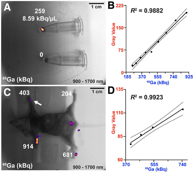
FIGURE 3.
In vitro and ex vivo SWIR CLI (900–1,700 nm) radioisotope sensitivity limit for 68Ga radiolabeled SiNPs. (A) SWIR CLI radioisotope in vitro detection limit for 68Ga-SiNPs after multiple half-lives. (B) SWIR CLI decay tracking to the limit of detection, linear regression (solid black line, R2 = 0.9882) and 95% CIs (solid gray lines) are shown. (C) Ex vivo SWIR CLI limit of detection for 68Ga-labeled SiNPs. The detection limit slightly worsens in tissue compared with in vitro imaging (∼140 kBq less sensitive). (D) Linear regression analysis (R2 = 0.9923) of ex vivo SWIR CLI of 68Ga-labeled SiNPs to limit of detection (403.3 kBq, C), 95% CIs are shown (solid gray lines). Detection limit paw is labeled with a white arrow.

