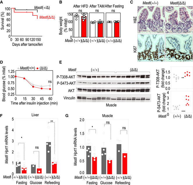
Survival curve of adult Mastl(+/Δ) (n = 11) and Mastl(Δ/Δ) (n = 14) mice after continuous tamoxifen treatment. No significant differences were found in the log‐rank (Mantel–Cox) test.
Plots representing the relative body weight of the mice included in the GTT assay from Fig
4. Body weight gain after 9 weeks of HFD (Left); body weight loss 1 week after tamoxifen injection (Middle); body weight loss after 16 h fasting (Right).
n = 6
Mastl(+/+) and
n = 11
Mastl(Δ/Δ). Error bars indicate SEM (unpaired student's
t‐test).
Representative images of the intestine from Mastl(+/+) and Mastl(Δ/Δ) mice showing the normal architecture of the epithelia by hematoxylin and eosin (H&E) staining (upper panel) and similar levels of proliferation (Ki67 staining, lower panels). Scale bars, 25 μm.
Insulin tolerance test (ITT) in Mastl(+/+) (n = 6) and Mastl(Δ/Δ) (n = 11) mice. Data are mean ± SEM; ns, not significant; two‐way ANOVA.
Immunoblot with the indicated antibodies in muscle tissues from Mastl(+/+) (n = 8) and Mastl(Δ/Δ) (n = 7) mice. Mice were fasted overnight for 16 h and sacrificed for sample collection. Quantification of the relative fold change signal of phospho‐AKT T308 and phospho‐AKT S473. Data are mean ± SEM; **P < 0.01; ***P < 0.001, unpaired Student's t‐test.
RT–qPCR analysis of Mastl mRNA in liver tissues from Mastl(+/+) and Mastl(Δ/Δ) mice fasted overnight for 16 (n = 7 mice/genotype), and injected intraperitoneally with glucose (2 g/kg body) (n = 3 mice/genotype), or re‐fed for 2 h and sacrificed 30 min later for sample collection refeeding conditions (n = 4 mice/genotype). Hprt1 was used as a housekeeping gene to normalize Mastl expression level. Plots show the mean + SEM; ns, not significant; *P < 0.05; **P < 0.01, unpaired Student's t‐test.
RT–qPCR analysis of Mastl mRNA in muscle tissues from Mastl(+/+) and Mastl(Δ/Δ) mice fasted overnight for 16 (n = 8 control mice and n = 5 Mastl KO mice), and injected intraperitoneally with glucose (2 g/kg body) (n = 4 control mice and n = 3 Mastl KO mice), or re‐fed for 2 h, and sacrificed 30 min later for sample collection refeeding conditions (n = 3 mice/genotype). Hprt1 was used as a housekeeping gene to normalize Mastl expression level. Plots show the mean + SEM; ns, not significant; *P < 0.05, unpaired Student's t‐test.

