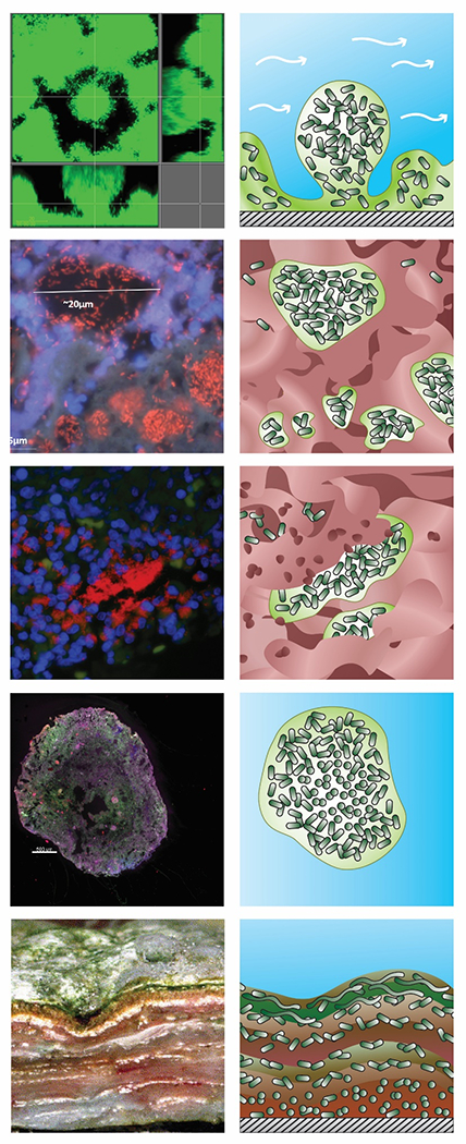
Figure 3.

Variety of biofilm structures underscoring differences between in vitro and in vivo or environmental biofilms. Original images are shown in the left column and a schematic drawing of the structure and its organization in the right column with shading denoting water (blue), aggregated microbial cells (dark green) and their extracellular polymeric substances (light green), host cells and other material including mucus or tissue (red), and attachment surface (hatched grey). A: Mushroom structure of Pseudomonas aeruginosa biofilm in vitro in a flow cell. B: Mucus embedded aggregates of P. aeruginosa surrounded by polymorphonuclear leukocytes in a cystic fibrosis lung (108) C: Wound-embedded aggregates of P. aeruginosa surrounded by polymorphonuclear leukocytes(109). D: Aerobic granules from a full-scale AquaNereda® wastewater treatment process (image courtesy of Kylie Bodle and Cat Kirkland). E: Striated microbial mat from a Brazilian lake(110). (Jill Story assisted with figure preparation).
