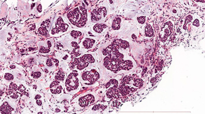
Fig. 1.
Liver metastasis core needle biopsy sample histology. Well-differentiated mucinous adenocarcinoma characterized by abundant pools of mucin and epithelial islands of atypical cells with ductal differentiation (HE staining, ×100 magnification, scale bar, 500 μm). The immunophenotype of tumor cells was GATA3+, TTF1−, CDX2−, ER+, PR−.

