Fig. 1.
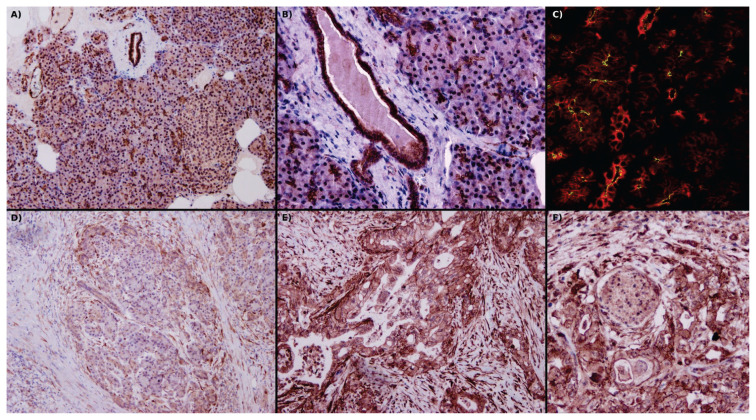
(A) Image of a physiological pancreas, with strong expression of PAR2 in the apical part of the acini and intralobular ducts, diffuse in interlobular ducts and the endothelium of vessels, and weak diffuse positivity in Langerhans islets. Fibroblasts are negative. (B) Detail view of PAR2 positivity in interlobular ducts, acini, and venules. (C) Apical positivity of PAR2 (green) on acini and interlobular duct cells, where cytokeratins 8/18 diffusely fills cytoplasm (red). (D) In pancreatitis, acini and intralobular duct cell PAR2 positivity decreases and takes on a diffuse cytoplasmic pattern. At the periphery, there is enhanced PAR2 positivity in fibroblasts and immune cells. (E) In pancreatic ductal adenocarcinoma, there is strong membranous positivity of PAR2 as well as greatly enhanced expression of PAR2 in fibroblasts. (F) Proximity of ductal adenocarcinoma to nerves results in enhanced neural PAR2 positivity. In A, B, D, E & F use immunohistochemical localization of PAR2, and in C, localization of PAR2 is by immunofluorescence in a confocal microscope. Original magnification 40× in A and D; 100× in B, E & F; 630× in C. P. Suhaj provided these archival pictures.

