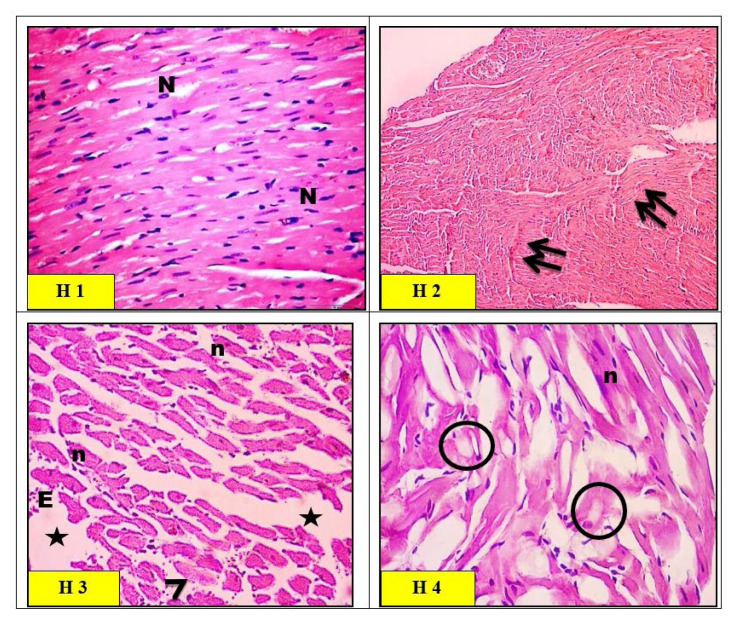
Fig. 8.
Light micrographs of left ventricular myocytes of heart in control group (H1) rats showing a normal histological pattern. The cardiac muscle fibers exhibited acidophilic sarcoplasm and ran in different directions with branching and anastomosing. The cardiac muscle nuclei were vesicular and centrally located (N). Light micrographs of zinc oxide nanoparticles (ZnO-nps) group (H2) rat heart showed thin distorted cardiac muscle fibers with areas of fragmentation and complete fiber loss (arrows). Waviness and hypereosinophilia of myofibers were also noticed (double arrows). Light micrographs of aluminum oxide nanoparticles (Al2O3-nps) group (H3) rat heart showed the interstitial spaces appearing wide (*) with some foci of extravasation (E). Some fibers exhibited pale sarcoplasm (arrowhead), and other fibers showed localized hypereosinophilia with pyknotic nuclei (n). Light micrographs of combination (ZnO-nps + Al2O3-nps) group (H4) rat heart showed widespread fragmentation and degeneration of cardiac muscle fibers. Large areas appear with pale vacuolar content (circles), and other areas of localized hypereosinophilic sarcoplasm with pyknotic nuclei (n) were also seen (H&E stain. Microscopic magnification ×400).

