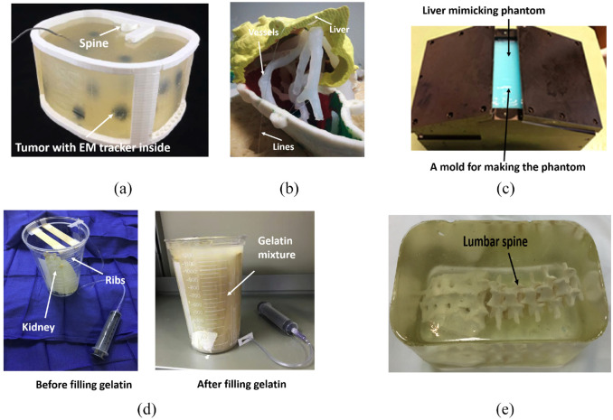
Figure 3.
(a) The developed abdominal phantom,20(b) Phantom liver assembly. The vessels and the lesions have been held in the correct position by means of lines,7 (c) The mold used to construct liver-mimicking phantom from PVA,21 (d) The $5 nephrostomy training phantom,22 and (e) The developed lumbar puncture training phantom.23

