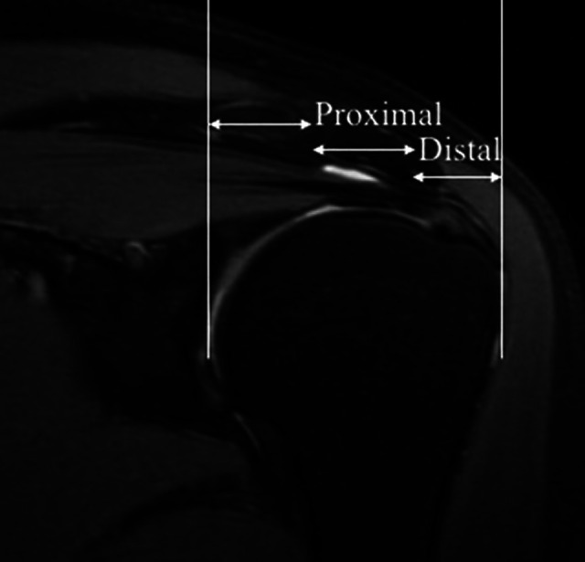
Figure 1.

Representative coronal oblique proton density–weighted fat-saturated image of tendon segmentation. The white lines represent landmarks that were manually chosen at the greater tuberosity and the articular surface of the humerus to divide the tendon into 3 equal parts: lateral (distal region); middle (proximal region); and medial (muscle region), which was not studied in the present investigation.
