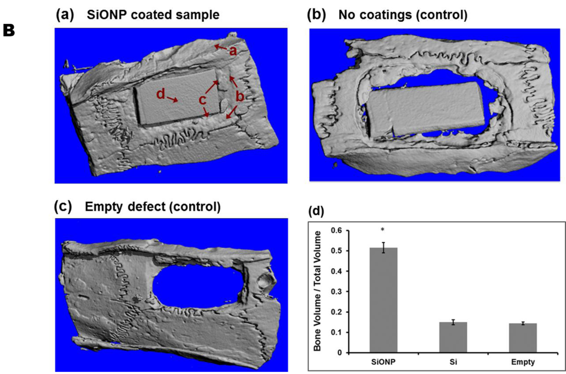
Figure 7.

MicroCT micrographs showed that (a) SiONP samples induced rapid bone-regeneration process and nearly completely filled interfacial gap within 5 weeks in-vivo whereas the control samples (b) and (c) didn’t show much bone to fill the gap over the same time period. (d) Quantitative analysis shows larger volume of mineralized bone regenerated for the SiONP surfaces, exhibited by larger bone to volume (B/V) ratio (5 folds) when compared to control surfaces. The arrows associated with a, b, c, and d in part (a) represent four different regions as we move from surrounding bone toward the center of the implant.
