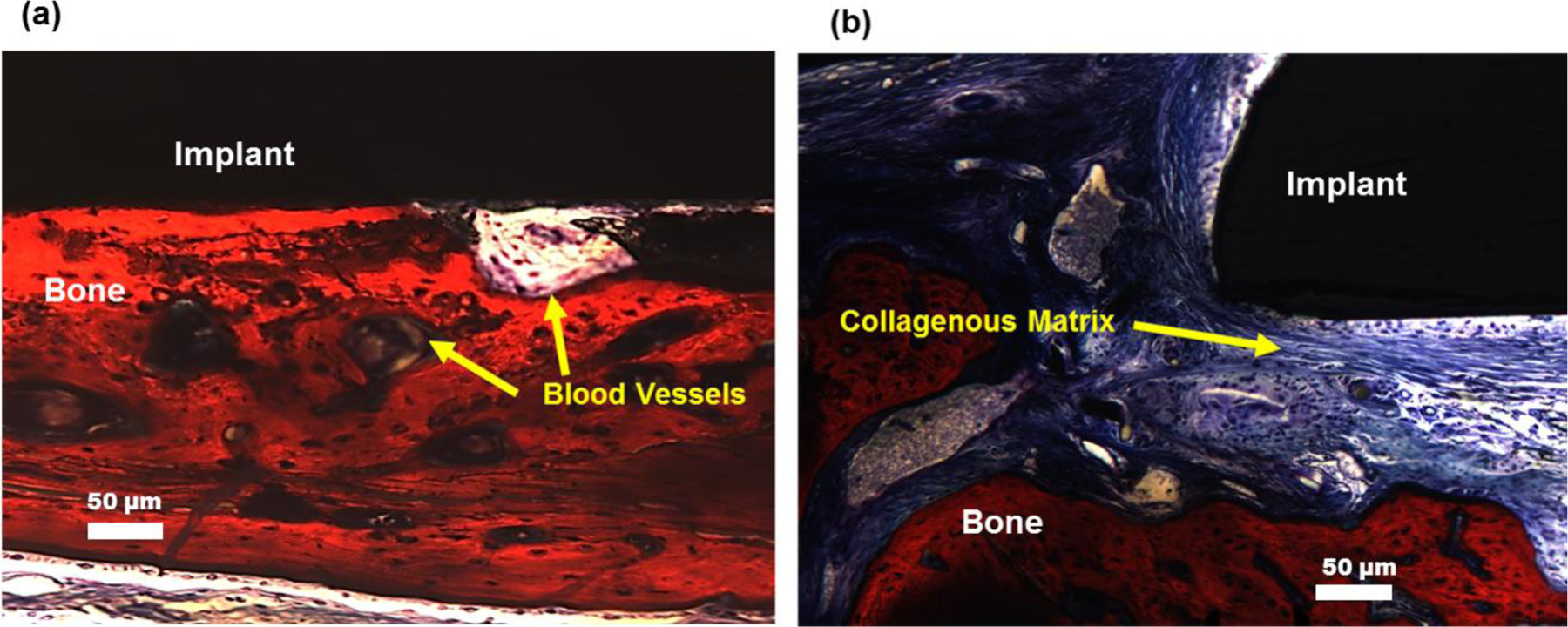
Figure 8.

Histology of extracted samples after 5 weeks of recovery demonstrate that (a) SiONP surfaces showed fully mineralized bone at the bone implant interface whereas (b) the control samples didn’t show much mineral formation but mostly collagenous fiber at the bone-implant interface.
