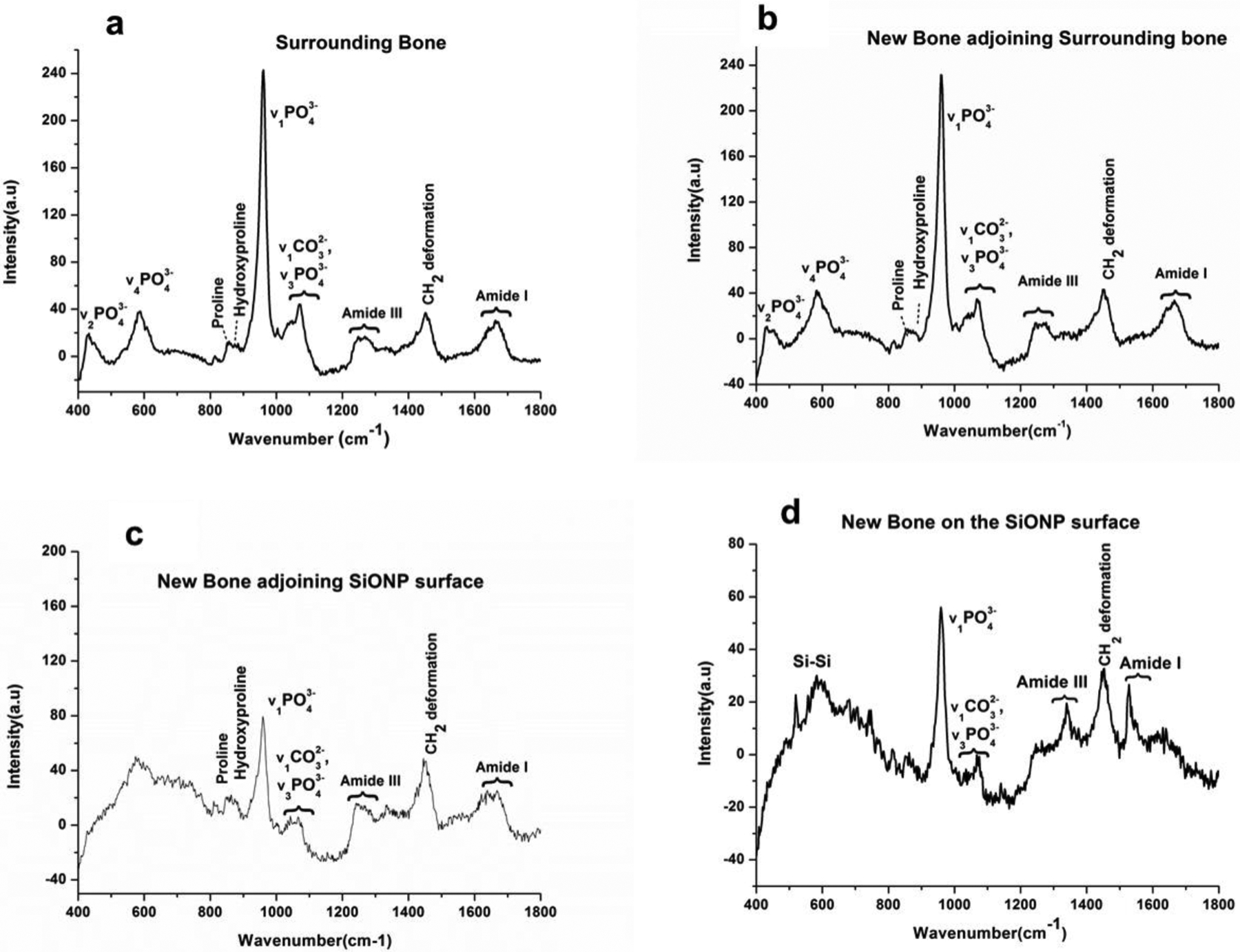
Figure 9.

Raman spectra compares the presence of mineralized content in the newly formed bone on the calvarial bone-implant extract surface as we move from surrounding (non-defected) bone towards the middle of the implant (SiONP). (a) Surrounding bone representing the non-defected original bone shows all the peaks expected to be present in a mature rat bone. (b) High intensity of v1PO43- peak indicates the complete mineralization of the newly formed bone at the interface of new/surrounding bone. (c) A drop in v1PO43- peak intensity and presence of distinct peaks for collagen, Amide III, Amide I and CH2 deformation indicate the presence of collagenous matrix and some immature mineralized content whereas (d) presence of peaks for Amide III, Amide I, CH2 deformation and poorly crystalline phosphate-carbonate are indicative of immature mineralization and/or P coordinated with O on SiONP surface. The regions a,b,c, and d are marked in Figure 7(a).
