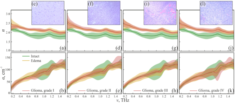
Fig. 12.
RI n, absorption coefficient α (by field), and representative H&E-stained histology of gelatin-embedded human brain gliomas of the different WHO grades ex vivo. (a)–(c) WHO Grade I. (d)–(f) WHO Grade II. (g)–(i) WHO Grade III. (j)–(l) WHO Grade IV (glioblastoma). Optical properties of tumors are compared with intact and edematous tissues. The error bars define a ± 2.0σ confidential interval of measurements (σ is the SD), which accounts for the optical properties variation within each tissue type. Reproduced from Ref. [147] published by SPIE under a Creative Commons Attribution 4.0 License.

