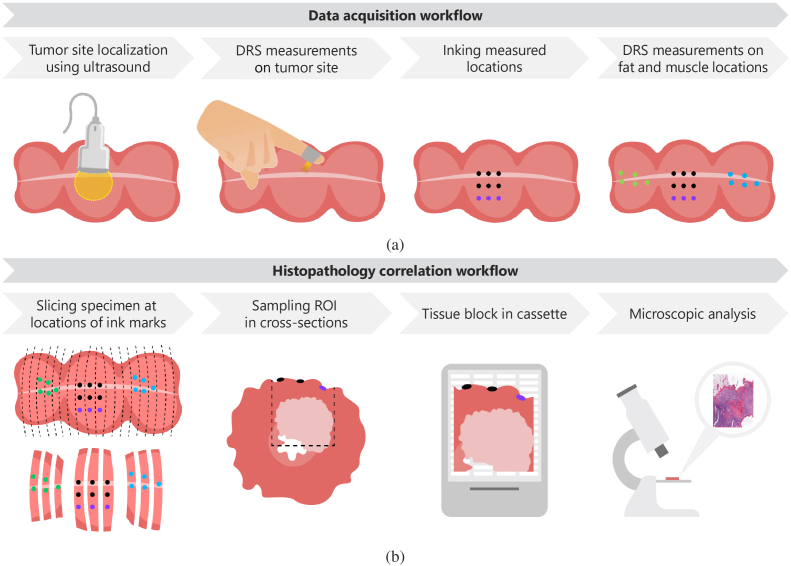
Fig. 5.
(a) Data acquisition workflow: First, the tumor site is localized in the freshly excised rectal specimen using ultrasound imaging (dashed yellow circle). In the next step, three to nine DRS measurements were performed using the side-firing fiber probe in this area, depending on the specimen size. Immediately after each measurement, the measurement location was marked with ink to allow correlation with histopathological results. The green and blue dots represent five DRS measurement locations on healthy muscle and fat tissue, respectively. (b) Histopathology correlation workflow: After finishing the data acquisition, the specimen was taken to the pathology department for further processing according to standard protocols. The specimen was fixated in formalin, after which it was dissected in slices. From these slices, the areas with ink marks on the surface were sampled in cassettes. The final H&E sections were digitally scanned for microscopic analysis and were annotated by a pathologist, who delineates fat, healthy rectal muscle, and tumor areas. The black and orange ink marks, representing the exact measurement locations, can be found back in the digital tissue slices as well as on the border of H&E images. In this way, tumor presence can be confirmed for every measurement location.

