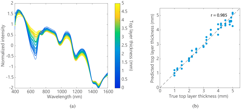
Fig. 10.
(a) DRS spectra of all measurement locations on the bilayer phantom, acquired using a side-firing fiber probe with a source-detector fiber distance of 2.7 mm (after SNV). The line color indicates the top layer thickness at the measured locations, which is equal to the distance to the Methylene blue layer (recognizable by the absorption dip at 664 nm), (b) Predicted top layer thickness versus true top layer thickness for all measurement locations on the bilayer phantom.

