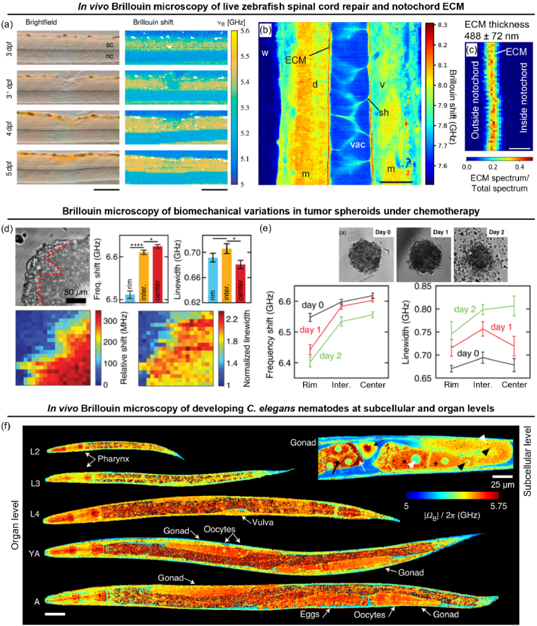
Fig. 8.
Imaging subcellular high-frequency biomechanics with BM. (a) BM of zebrafish larvae captures mechanical changes before (3 dpf), after spinal cord injury (3+ dpf), and post-repair (4-5 dpf). Scale bar: 150 µm. dpf: days post fertilization, Sc: spinal cord, nc: notochord. Reprinted from [17], with permission from Elsevier. (b) High-resolution BM of zebrafish notochord combined with double-peak spectral fitting resolves (c) sub-micron notochord ECM thickness. Scale bar: (b) 20 µm and (c) 1 µm. d: dorsal, v: ventral, m: muscle, sh: sheath cell, vac: vacuole. (b) and (c) Adapted with permission from [14] © Optica. (d) Phase contrast (top) and BM maps (bottom) of elasticity (frequency shift, left) and viscosity (linewidth, right) in live tumor spheroid, revealing stiffer elastic center (red bars) and softer viscous outer rim (blue bars). (e) Mechanical changes in the spheroid over 2 days of chemotherapy; phase contrast (top) shows disaggregation at the outer rim by Day 2. (d) and (e) Reprinted from [111] with permission. Copyright 2019 by the American Physical Society. (f) Mechanical changes (frequency shift, ΩB) in a developing nematode (larvae stages L2, L3, L4, young adult YA, and adult A) at the organ level. High-resolution BM of an adult gonad at the subcellular level (inset) reveals nuclei and nucleoli (white and black arrowheads), cytoplasm (asterisk), uterus edge (white arrow), oocyte pushed from the spermatheca into the uterus (black arrow), and a four-cell embryo in the uterus (black diamond arrow). Adapted by permission from Springer Nature: [16].

