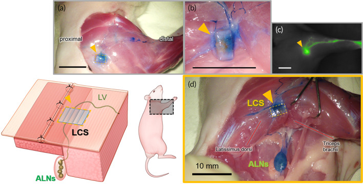
FIGURE 1.

After 8 weeks of implantation, (a) the lymphatic flow through the lymphatic channel sheet (LCS) (yellow triangle) of polydimethylsiloxane (PDMS) from the distal to the proximal area could be identified by the Evans blue (EB) dye. (b) It is seen that the lymphatic vessels were connected at both ends of the LCS after removing the surrounding tissue in the magnified view. (c) The indocyanine green (ICG) fluorescence image of the lymphatic flow near the LCS. The lymphatic fluid was collected from the LCS. (d) The anatomical detail of the lymphatic flow from nearby the LCS to axillary lymph nodes (ALNs). The EB dye, which was injected into the palm, flowed into ALNs throughout the LCS. The pectoralis major was incised to identify lymph vessels from the distal area to ALNs and surrounding the LCS. All figures were from the same animal and all scale bars are 10 mm.
