
FIGURE 6.
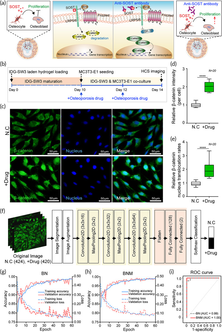
Osteoporosis drug testing using bone‐on‐a‐chip. (a) A schematic showing the Wnt pathway related to bone formation, one of the target mechanisms for osteoporosis treatment. SOST, secreted by osteocytes, downregulates osteoblast proliferation (left image) whereas an anti‐SOST antibody (used as an osteoporosis drug) upregulates osteoblast proliferation through nuclear translocation of β‐catenin via the Wnt pathway (right image). (b) The overall sequential process for osteoporosis drug treatment in the bone‐on‐a‐chip. After 10 days of IDG‐SW3 maturation for SOST secretion, MC3T3‐E1 cells were cocultured for additional 4 days. Cells were treated twice with an osteoporosis drug on Days 10 and 12. (c) Representative images of MC3T3‐E1 cells in osteoporosis drug‐treated and untreated groups that were immunostained against β‐catenin (green) and nucleus (blue). (d) Relative average fluorescence intensity of β‐catenin in cells in each drug‐treated group compared to that in the control (N = 20). (e) Relative β‐catenin nuclear translocation rate in drug‐treated group compared to that in the control (N = 20). (f) The proposed CNN‐based deep learning architecture to perform osteoporosis drug testing. (g and h) Accuracy (left x‐axis) and loss curves (right x‐axis) of CNN training and validation for the (g) BN and (h) BNM datasets with successive epochs. (i) ROC curve analysis comparing classification results in BN and BNM datasets. AUC, area under the ROC curve; BN, β‐catenin and nucleus fluorescence image data; BNM, β‐catenin, nucleus, and their merged image data; CNN, convolutional neural network; ROC, receiver operating characteristic