
FIGURE 1.
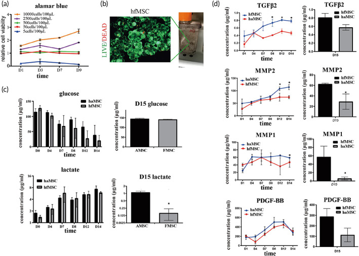
Large scale expansion of human fetal mesenchymal stem cells (hfMSCs) in a vertical‐wheel bioreactor. (a) Cell viability of various density of cells was measured using Alamar blue test. The data were normalized to negative control of all time points. Starting cell density was 1 × 10⁶ cells/100 ml. N = 3; (b). Live/dead cell staining results showed that fetal MSCs and microcarriers formed aggregates and more than 90% of the cells were alive at Day 14 culture; (c). Cell metabolic index changes during culture. The glucose concentration kept on decreasing during culture, indicating that cells were actively proliferating. After change into serum‐free media, at Day 15, lactate concentration was lower in the hfMSCs culture suggesting hfMSC have a unique pattern of energy metabolism, N = 3; (d). During 14 days culture, ELISA results showed that the concentration of TGFβ2, VEGF, MMP1, and MMP2 were significantly higher in media of hfMSCs than those in haMSCs media; Statistic: Mann–Whitney U (unpaired, nonparametric T‐tests). N = 3, *p < 0.5. Brightness and contrast were adjusted for clear demonstrated contents of the representative pictures. TGFβ2, Transforming growth factor beta‐2; VEGF, Vascular endothelial growth factor; MMP, matrix metalloproteinase.