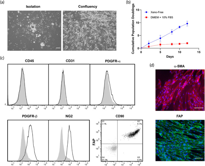
FIGURE 3.

Phenotypic characterization of human placental PCs under xeno‐free conditions. (a) Live phasecontrast microscopy images of a vessel fragment on a fibronectin‐coated plate after isolation from placental tissue and atthe confluency state. (b) The cumulative population doublings of PCs cultured under xeno‐free conditions is higher than in DMEM supplemented with 10% FBS. (c) Flow cytometry analysis of PCs cultured under xeno‐free conditions confirmed the expression of PDGFR‐α, NG2, CD90, and FAP. Furthermore, these cells lack expression of CD31 and CD45. (d) PCs showed positive expression of α‐SMA and FAP, as confirmed by immunofluorescence staining. Scale bar = 100 μm. Representative of three independent donors. DMEM, Dulbecco's modified Eagle medium; FBS, fetal bovine serum; PC, pericyte
