FIGURE 5.

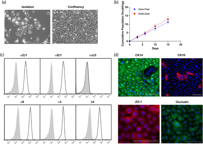
Phenotyping characterization of human keratinocytes under xeno‐free conditions. (a) Live phasecontrast microscopy images of keratinocytes after isolation from the epidermis of donor foreskin and at the confluency state. (b) The cumulative population doublings of keratinocytes cultured under xeno‐free conditions is comparable to KGM‐Gold medium. (c) Flow cytometry analysis confirmed the expression of integrins α2β1, α5β1, α6, α3, and β4, but not αvβ3. (d) Confocal microscopy exhibiting CK14, CK10, junctional ZO‐1, and intracellular occludin staining. Scale bar = 100 μm. Representative of three independent donors
