
FIGURE 6.
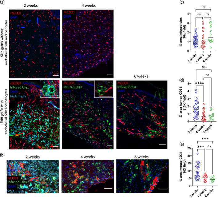
Characterization of graft‐derived and host‐derived microvessel formation in xeno‐free 3D bioprinted skin grafts with and without ECs and PCs 2‐, 4‐ and 6‐week postengraftment onto immunodeficient mice. (a) Fluorescent mouse CD31 staining depicts higher degree of host angiogenesis (red) and the presence of perfused human EC‐lined vessels (infused Ulex stain) in grafts containing ECs and PCs. (b) Human CD31 staining shows the presence of nonperfused human ECs (red) surrounded by PGA mesh and perfused human vessels (green). Nuclei are stained blue. Scale bar = 100 μm. Quantification of area of (c) infused ulex, (d) human CD31, and (e) mouse CD31 at 2‐, 4‐, and 6‐week postimplantation. Data are shown for three independent experiments. (*p < 0.05, **p < 0.01, ***p < 0.001, ****p < 0.0001, ns p > 0.05; one‐way ANOVA method with Tukey post hoc comparisons). 3D, three‐dimensional; ANOVA, analysis of variance; EC, endothelial cell; PC, pericyte; PGA, polyglycolic acid