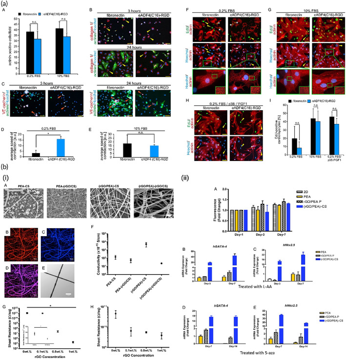
FIGURE 15.

Reduced graphene oxide application in cardiac tissue repair. (a) (A–C) Endothelial, Fibroblast, and CM cells attachments on the eADF4(C16)‐RGD coatings after three‐time points of 3, 24, and 48 h of incubation. (D,E) Quantitative analysis of contraction speed in the presence of 0.2% FBS and 10% FBS, respectively. (F–I) The CM cells were cultured on the eADF4(C16)‐RGD‐based nanostructures for investigating the proliferation and differentiation of the sarcomeres. Reproduced from Reference 318 with permission from Nature. (a) rGO‐poly(ester amide) conductive scaffolds and their potential for cardiac tissue repair. (A) SEM images demonstrating fiber morphology and fiber diameter distribution. (B–D) (rGO/PEA)‐CS (with rGO in the core) where the core is labeled with red DiI (B) and the shell auto‐fluoresces blue (C), and the purple overlap (D) demonstrating the mixing of the core and shell and subsequent lack of clear core‐shell morphology. (E) TEM image indicating non‐homogenous mixing and lack of core‐shell structure. (F) Conductivity of different three‐component coaxial scaffolds. (G,H) The effect of rGO concentration on film resistance of Composite rGO/PEA and rGO/CS films, respectively. (I) Cell proliferation and differentiation on PEA, rGO/PEA P, and (rGO/PEA)‐CS scaffolds. (J–M) iPSC‐derived MSC gene expression. Reproduced from Reference 319 with permission from Elsevier
