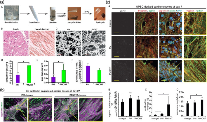
FIGURE 16.

Carbon nanotube adorned hydrogel for cardiac tissue engineering. (a) (A) Illustration of different steps in preparation of pericardial matrix (PM)‐ and PMCNT‐gels. (B) hematoxylin–eosin staining histological images of the fresh and decellularized pericardium. (C) SEM images of PM‐ and PMCNT‐gels. (D–F) Quantitative analyses of electrical conductivity, pore diameter, and swelling ratio, respectively. (b) confocal images of cell‐laden tissue construct stained for the CM‐specific markers troponin I and sarcomeric‐α‐actinin. (c) Fabricated hydrogels intrinsically increase intercellular electrical coupling of hiPSC‐derived CMs. Reproduced from Reference 341 with permission from The Royal Society of Chemistry
