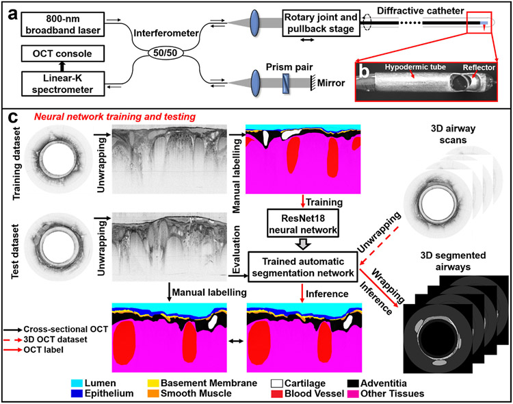
Fig. 1. 800-nm diffractive OCT system and workflow for neural network training and testing.
(a) Schematic of the diffractive spectral-domain OCT system operating at 800 nm with a customized linear-K spectrometer and an achromatic diffractive catheter of a 1.3-mm diameter. (b) Photo of the distal end of a diffractive catheter encased within a hypodermic tube. (c) For neural network training, unwrapped OCT images in the training dataset were manually labeled by an experience OCT reviewer. After that, the trained network first underwent the performance evaluation on the test dataset, the segmentation results were compared with the manually labeled ground truth. Then, the trained network was tested on 3D airway scans to automatically recognize and segment airway microstructures for volumetric visualization and quantification of small airways of sheep.

