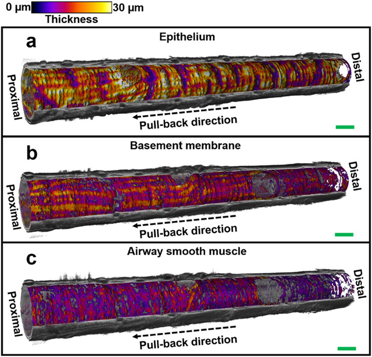
Fig. 5. 3D visualization of small airway tissue compartments.
Visualization of the volumetric architectures of tissue compartments (embedded in the cut-away view of the reconstructed 3D OCT image) in (n=1) 18-mm long sheep small airway, including epithelium (a), basement membrane (b), and airway smooth muscle (c). Thickness of each tissue compartment is represented with color and the cut-away view of 3D OCT image is represented with gray scale. Scale bars: 1 mm.

