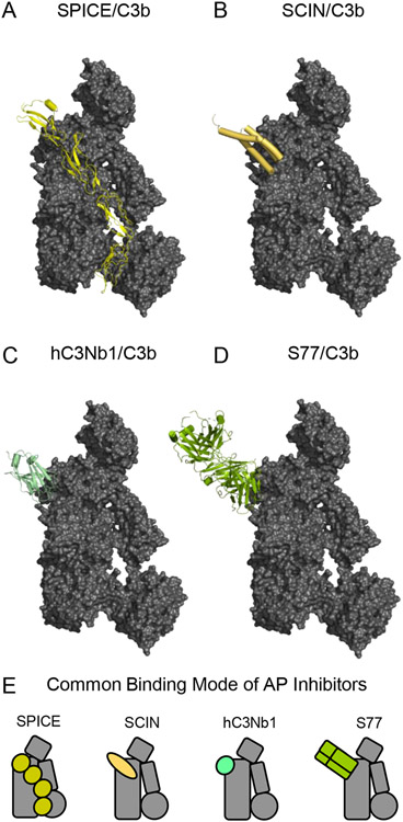
Figure 5.
Structural Features of C3b-binding Inhibitors of the Alternative Complement Pathway. (A) Structure of C3b bound to CCP domains 1-4 of the Variola virus complement inhibitor SPICE as drawn from PDB entry 5FOB [43]. C3b is colored as a grey surface, while SPICE is colored yellow. (B) Structure of C3b bound to the S. aureus immune evasion protein SCIN as drawn from PDB entry 3OHX [22]. C3b is colored as a grey surface, while SCIN is colored pale orange. (C) Structure of C3b bound to the nanobody hC3Nb1 as drawn from PDB entry 6EHG [67]. C3b is colored as a grey surface, while hC3Nb1 is colored light green. (D) Structure of C3b bound to the F(ab) fragment of monoclonal antibody S77 as drawn from PDB entry 3G6J [68]. C3b is colored as a grey surface, while S77 is colored olive green. (E) Simplified representations of the structures shown in panels A-D. Note the common binding surface recognized by each of these C3b-binding Alternative Pathway inhibitors.

