Figure 6.
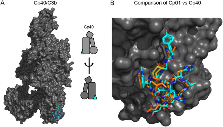
Structural and Functional Features of Compstatin Class Complement Inhibitors. (A) Structure of C3 bound to the third-generation Compstatin Cp40 as drawn from the PDB entry 7BAG [74]. C3b is colored as a grey surface, while Cp40 is shown in stick convention and colored blue. A simplified representation of the structure is inset at the right of the panel. (B) Comparison of bound structures for the second-generation Compstatin Cp01 and Cp40 as drawn from the PDB entries 2QKI [75] and 7BAG [74], respectively. Both inhibitors are drawn in stick convention with the carbon atoms of Cp01 in orange and those of Cp40 in blue. Note that this image was constructed by superimposing the structure of Cp01 bound to C3c upon that of Cp40 bound to C3b using the keyring core of C3 as the basis for calculations.

