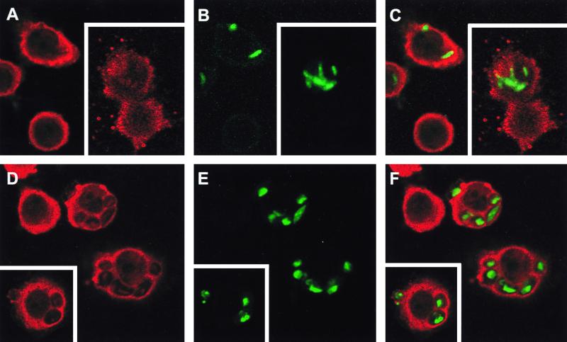
FIG. 6.
Analysis of NADPH oxidase assembly at the phagosomal membrane by confocal microscopy. Phagocytosis of FITC-labeled M. kansasii (A to C) or FITC-labeled zymosan (D to F) was not synchronized in order to obtain phagosomes at different maturation stages. Following phagocytosis, differentiated U937 cells were fixed, permeabilized, and stained with anti-p47phox antibodies revealed by TRITC-conjugated anti-rabbit antibodies. The stained cells are representative of three independent experiments, and phagosomes visualized by confocal microscopy are shown. (A and D) p47phox staining alone; (B and E) FITC-labeled particles alone; (C and F) superimposition of both types of staining. Insets, representative of a second experiment.

