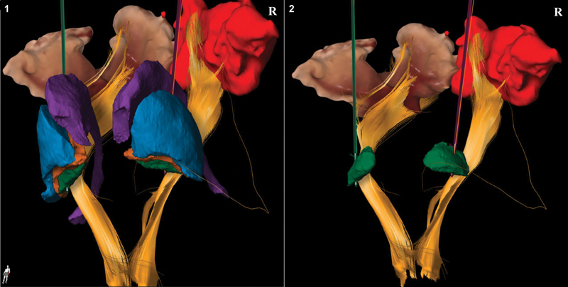
Fig. 1.

Three-dimensional representation of intraoperative placement of deep brain stimulation (DBS) leads in patient 2. Different colors are used to represent the anatomical structures adjacent to the globus pallidus interna (GPi) for DBS lead placement. The lead in green indicated the left lead, whereas the lead in purple depicts the right lead. Left panel: anterior view of the basal ganglia and corticospinal tracts. The following structures are depicted: GPi (green), globus pallidus externa (orange), putamen/ lentiform nucleus (blue), caudate nucleus (purple), corticospinal tracts (yellow). Right panel: location of the DBS leads in the GPi (green) with adjacent structures removed.
