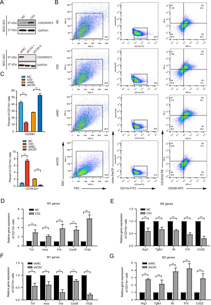
Fig. 2. The xenograft tumour model proves that the high expression of CDK5RAP3 in gastric cancer inhibits the polarization of macrophages towards the M2 type.
A Western blotting was used to detect the protein expression of CDK5RAP3 in the protein extract of the gastric cancer cell line BGC-823. B Flow cytometry was used to detect the expression of CD16/32 and CD206 on the surface of CD11b + macrophages and to determine the percentage of CD16/32+ and CD206+ cells in CD11b + macrophages. C The bar graphs present the difference of Q1 quadrant (representing the percentage of CD16/32-PE staining positive cells) and Q3 quadrant (representing the percentage of CD206-APC staining positive cells) in different treatment groups in FACS plots. D, E Relative gene expression of M1 markers (Tnf, Inos, Il1b, Cxcl9 and Il12a) and M2 markers (Arg1, Tgfb1, Vegfa, Il6, Il10 and Ccl22) (F, G) in the tumour tissues of mice. *P < 0.05; **P < 0.01.

