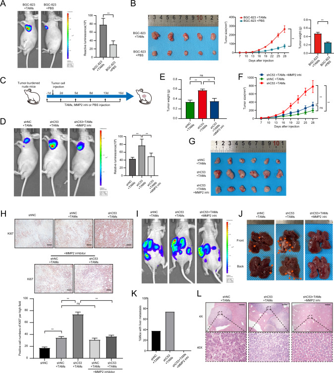
Fig. 7. The absence of CDK5RAP3 in gastric cancer cells mediates the cancer-promoting phenotype of macrophages in a xenograft tumour model.
A Representative bioluminescence images of mice 4 weeks after subcutaneous injection of BGC-823 cells and/or macrophages, and the images were quantified. Representative photos and tumour growth of each group of mouse tumours are shown in (B). The size of the xenograft was measured every 3 days until the mice were sacrificed. The average tumour weights of the three different groups were compared. (n = 5 per group). C MMP2 inhibitors were applied to the experimental design of BALB/c nude mice in xenograft tumour models. D Representative bioluminescence images of mice 4 weeks after subcutaneous injection of gastric cancer cells and TAMs, and the images were quantified. E The average tumour weights of the three different groups were compared. F The size of the xenograft was measured every 3 days until the mice were sacrificed. G Representative pictures of tumours in each group of mice are shown. (n = 6 per group). H Ki67 pathological images of tumours collected from Balb/c nude mice injected with BGC-823 cells and/or TAMs. IHC staining of Ki67 in tumour tissues in mice xenograft model and positive cell numbers per high field were counted. Scale bar = 50 μm. I Representative bioluminescence images of mice 4 weeks after spleen injection of BGC-823 cells and TAMs, and the images were quantified. J Representative images of liver metastasis. K The percentage of tumour metastasis in the Lenti-scr + TAMs groups, Lenti-shC53 + TAMs/PBS groups and Lenti-shC53 + TAMs/MMP2 inhi groups (n = 8 per group). L Representative haematoxylin-eosin-stained sections of liver metastases in mice 28 days after implantation in different groups. Magnification: ×4 and ×40. Scale bar = 200 µm. *P < 0.05; **P < 0.01.

